
In the past twenty years, the healthcare sector has is revolutionised with technology. MRI, CT scan and X-ray are some of the excellent techniques that are employed for quick diagnosis of diseases. In fact if we have to discern the present scenario, an ideal way to detect the COVID 19 symptoms would be with the help of chest X-rays. Though these methods entail an enormous amount of investment, they serve the purpose.
However, one of the drawbacks while using this technique is that they are time-consuming. MRIs which are deemed to be the best tool for diagnosing diseases and conditions can take up to 4-5 hours to generate results. It becomes an exhausting experience for the patients to sit for long hours, and especially for young children, who often find it challenging to adjust their bodily posture with this technology.
That's why, to counter this problem, the researchers at Facebook and NYU have collaborated in project, FastMRI initiative, to use Artificial Intelligence for advancing up MRI scans for more accelerated results, without compromising the quality of the generated image.
Magnetic Resonance Imaging uses a large magnetic radio wave to create a detailed cross-sectional image of internal organs and structures. An MRI scanner contains two powerful magnets.
The human body is made up of water molecules, which contains hydrogen and oxygen atoms. At the core of these atoms a small particle, Proton serves as a magnet for any magnetic field. When the first magnetic field is deployed, the water molecules get aligned in either north or south direction. The second magnetic field generates a series of pulses by turning on and off, causing hydrogen atom to change its alignment when the magnetic field is on and to resume back to its original relaxed state when switched off.
The electricity is then passed through the gradient coil by vibrating creates a magnetic field/signal, which is responsible for the cracking sound inside the scanner. These signals are collected by the scanner as a sequence of individual 2D frequency measure known as K-space data.
After entire data is collected, the system then applies a complex mathematical formula- inverse Fourier transforms, which would create a detailed MRI of the associated body part with the help of raw K space data.
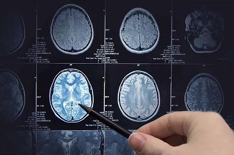
The magnetic field can then aid in producing cross-sectional images of any part of the body. It is used for detecting the anomalies of the brain and spinal cord, tumours, cysts, breast cancer, knee or joint injuries, heart and liver anomalies and others.
The fastAI team built a neural network for training to create a large number of data sets. For this process, the data set of 108 MRI scans with different diseases and conditions were analysed. In each scan, three-fourth of the data was removed and fed to the AI model. It was observed that with the use of this AI-model, the scans that were generated were four times faster than the traditional scans. The image generated with this model was also distinguishable from that of the traditional scan.
No evident ghostly images, which are generated when the patients are unable to hold themselves in a stagnant position during MRI, was observed while using this model.
The researchers are now planning to use this technology for doing scans and generating scans in other body parts such as the brain. A detailed study about the application of this new technology is produced in the American Journal of Roentgenology.
The Future Prospects of AI-based MRI
The future with this AI-model is vast. The healthcare burden would be lesser by applying this model.
Join our WhatsApp Channel to get the latest news, exclusives and videos on WhatsApp
_____________
Disclaimer: Analytics Insight does not provide financial advice or guidance on cryptocurrencies and stocks. Also note that the cryptocurrencies mentioned/listed on the website could potentially be risky, i.e. designed to induce you to invest financial resources that may be lost forever and not be recoverable once investments are made. This article is provided for informational purposes and does not constitute investment advice. You are responsible for conducting your own research (DYOR) before making any investments. Read more about the financial risks involved here.